Northeastern University & Stanford University: Nano CuS system promotes photothermal treatment of deep tumors
Deep-Tissue Photothermal Therapy Using Laser Illumination at NIR-IIa WindowXunzhi Wu, Yongkuan Suo, Hui Shi, Ruiqi Liu, Fengxia Wu, Tingzhong Wang, Lina Ma, Hongguang Liu, Zhen Cheng Nano-Micro Lett. (2020)12:38 https: //doi.org/10.1007/s40820-020-0378-6
Highlights of this article
1 Synthesized polyethylene glycol-coated copper sulfide nanoparticles with similar absorption values at 808 nm and 1275 nm, and compared the capabilities of these two lasers in deep tissue photothermal treatment. 2 When using 5 mm pork tissue to shield tumor tissue to simulate the deep tissue environment, the 1275 nm laser exhibits a more excellent tumor photothermal treatment effect than the 808 nm laser.
brief introduction
Professor Liu Hongguang’s project from the Institute of Molecular Medicine, School of Life Sciences and Health, Northeastern University, was combined into polyethylene glycol-coated copper sulfide nanoparticles (CuS-PEG NPs) with similar absorption values at 808 nm and 1275 nm. The platform compares the capabilities of 808 nm and 1275 nm lasers in deep tissue photothermal treatment. In the comparative experiment, the 1275 nm laser showed a higher tissue penetration ratio, higher deep tissue heating ability and more effective deep tissue tumor killing ability than 808 nm laser. The photothermal therapy experiment carried out after the mouse tail vein injection of CuS-PEG NPs verified the feasibility of applying 1275 nm laser to photothermal therapy.
Graphic guide
I Synthesis and characterization of CuS NPs and CuS-PEG NPs
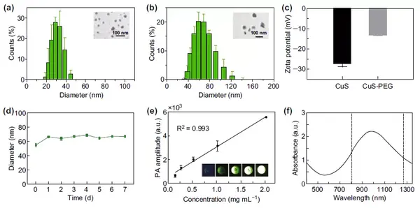
The CuS NPs were synthesized and PEG was further attached to the surface. Before and after connection, the hydrated particle size of the nanoparticles increased by 19 nm, and the surface potential increased by 14 mV. CuS-PEG NPs have good stability in serum, and the hydrated particle size has not changed significantly within 7 days. Within the test concentration range, the photoacoustic signal intensity of CuS-PEG NPs is proportional to its concentration. The absorption values of CuS-PEG NPs at 808 nm and 1275 nm are relatively close.

Figure 1 (a) CuS NPs hydrated particle size and transmission electron microscope image; (b) CuS-PEG NPs hydrated particle size and transmission electron microscope image; (c) CuSNPs before and after connecting with PEG surface potential changes; (d) CuS-PEG NPs Serum stability analysis; (e) CuS-PEG NPs photoacoustic signal intensity and its concentration relationship; (f) CuS-PEG NPs absorption spectrum. Comparison of the in vitro tissue penetration capabilities of II808 nm and 1275 nm lasers Firstly, the penetration capabilities of the two lasers for pork tissues of different thicknesses were compared. Under different thicknesses of pork tissues, the penetration ratio of 1275 nm laser is higher than that of 808 nm laser. Subsequently, the tissue is irradiated with the two lasers to raise the temperature, reflecting the difference in the scattering effects of the tissues on the two lasers. The scattering effect of tissue for 1275 nm laser is lower than that of 808 nm laser.

Figure 2 (a) The ratio of the remaining laser intensity after 808 nm and 1275 nm lasers penetrated pork tissues with different thicknesses; (b) The thermal image of two types of lasers irradiating pork tissues; (c) The sagittal and horizontal planes of pork loins The relative area where the upper temperature is higher than a certain value.
III Comparison of the deep tissue heating capabilities of the two lasers
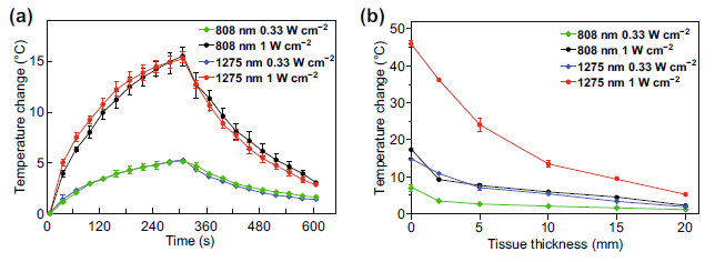
The two lasers with the same power density irradiated the CuS-PEGNPs solution with the corresponding concentration, and the CuS-PEGNPs increased the temperature closer. After covering pork tissues with different thicknesses to simulate the deep environment, the temperature of the solution in the 1275nm laser group was significantly higher than that of the 808nm laser group.

Figure 3 (a) CuS-PEG NPs heating curve under different laser conditions without tissue shielding; (b) CuS-PEGNPs solution heating curve under different thickness tissues under different laser conditions.
IV Comparison of the in vitro cell killing ability of the two lasers
4T-1 cells were plated on a 96-well plate, and different condition groups were set to test the killing ability of the two lasers on the cells. After the experiment, the cell viability was measured by MTT method, and the cell status was observed by Hoechst 33342 and PI double staining. The 1275 nm laser shows a stronger ability to kill deep cells than the 808 nm laser.

Figure 4 (a) MTT results of in vitro cell photothermal experiments with different thicknesses of pork tissues; (b) Hoechst-PI fluorescence double staining results after cell photothermal experiments.
V Comparison of in vivo tumor killing ability of two lasers
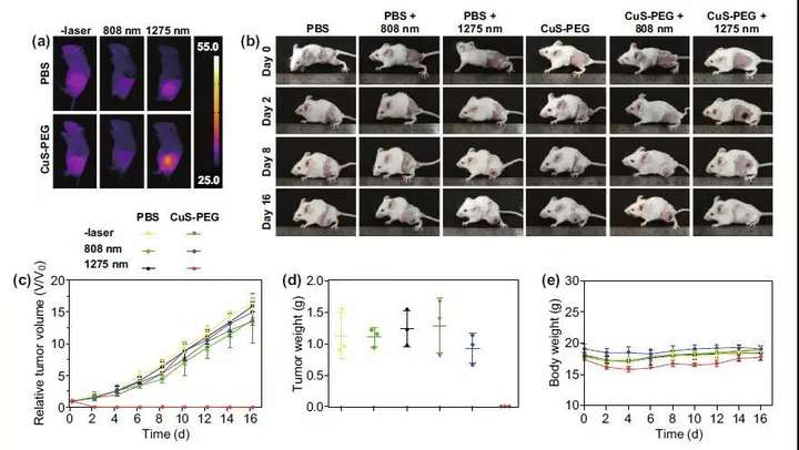
The tumor-bearing mouse model was constructed by subcutaneously inoculating 4T-1 cells. CuS-PEGNPs or PBS was injected into the tumor to cover 5 mm thick pork tissue, and then two lasers were irradiated. At the end of the experiment, the whole body thermal image of the mouse was recorded, and the whole body image, tumor volume and body weight of the mouse were recorded continuously for the next 16 days. On day 16, the weight of the tumor was recorded. Intratumoral injection of CuS-PEGNPs and irradiated with 1275nm laser showed good tumor photothermal treatment effect, but 808nm laser failed to effectively kill tumor tissue.

Figure 5 (a) Whole body thermal image of mice in different groups at the end of photothermal treatment; (b) Whole body images of mice before and after photothermal treatment; (c) Curve of relative tumor volume change after photothermal treatment; (d) photothermal treatment Tumor phase weight on the 16th day after the end of the treatment; (e) The weight change curve of the mice after the end of photothermal treatment.
This information is sourced from the Internet for academic exchanges. If there is any infringement, please contact us to delete it immediately
This information is sourced from the Internet for academic exchanges. If there is any infringement, please contact us to delete it immediately
18915694570
Previous: Chen Xiaoyuan/Yang Zhe


