Explore the unknown, the medical application potential of MOFs stimulated by nano

Renjun Pei, a research team from Suzhou Institute of Nanosaures, Chinese Academy of Sciences, systematically studied the design, synthesis and generation mechanism of porphyrin-based MOFs as well as their application in photodynamic therapy of tumors. The team found that the central metal coordination of the inner ring of Porphyrin (TCPP) plays a non-negligible role in the design and synthesis of MOFs, and the central metal coordination of the porphyrin inner ring can greatly affect the morphology and chemical properties of MOFs materials.
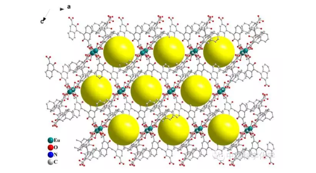
First, they constructed porphyrin gD-TCPp MOFs nanocrystals without central metal coordination (FIG. 1A) and demonstrated their high singlet oxygen yield. However, after the central coordination in the inner ring of porphyrins, even hydrogen protons can significantly change the MOFs morphology and uV-vis absorption (FIG. 1B and 1C). Based on these results, the effect of the central metal coordination of porphyrins was preliminarily proposed.

FIG. 1. (a) GD-TCPP and (b) SEM images of GD-(H2TCPP)2+, and (c) UV-visible absorption.
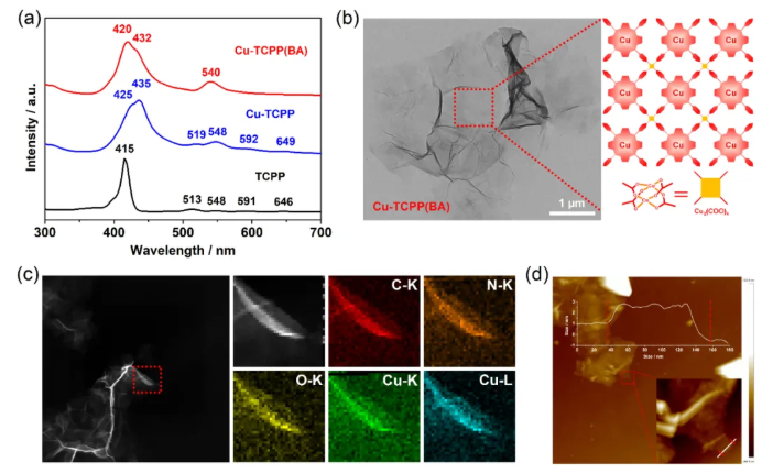
On this basis, the team designed and synthesized the porphyrin Cu-TCPp (BA) MOFs micron slices with central copper metal coordination with the assistance of benzoic acid

(FIG. 2), confirming that such porphyrins with central metal coordination can reduce the H accumulation and J aggregation between layers and cause the anisotropic growth of MOFs. This study revealed the growth mechanism of porphyrins, elaborated on the role of central metal coordination of porphyrins, and provided a feasible strategy for the synthesis of ultra-thin MOF materials with micron size.
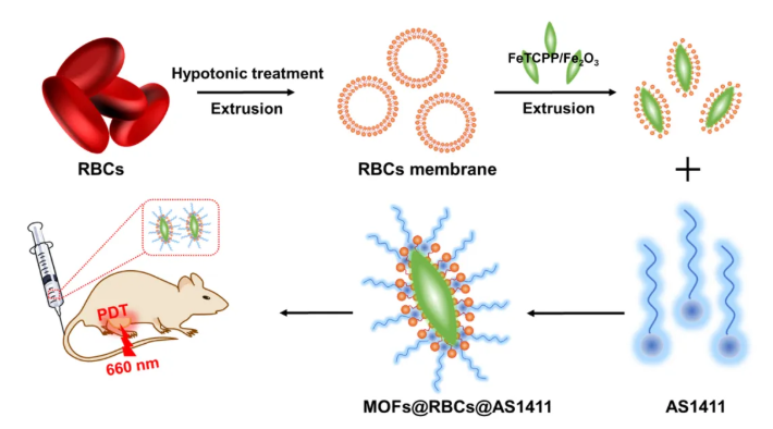
RBCs membrane was used to camouflage MOFs, improve blood circulation and tissue residence time in vivo, and AS1411 aptamer was modified to achieve high CONCENTRATION of MOFs in the tumor area (Figure 3). The encapsulation of RBCs membrane and the targeting effect of AS1411 aptamer enhanced the enrichment ability of MOFs nanoparticles in tumor sites, improved the PDT effect, and reduced the side effects. This study provides a new idea for promoting the application of MOFs nanomaterials in biomedical field.

FIG. 3 RBCs membrane encapsulation of FeTCPP/Fe2O3 MOFs and modification of AS1411 aptamer.
These work revealed the partial influence of the central metal coordination of the porphyrin inner ring on the MOFs structure, further elaborated the mechanism of MOFs material generation, provided some methods for designing and synthesizing MOFs materials, and proposed new strategies for improving the effect of photodynamic therapy on tumors.
Source: Suzhou Incubator Center, Chinese Academy of Sciences
This information is from the Internet for academic exchange only. If there is any infringement, please contact us to delete it immediately
18915694570
Previous: In-situ polymerization


