
Angiosteosis-Hydroxyapatite
Nano-hydroxyapatite accelerates vascular calcification via lysosome impairment and autophagy dysfunction in smooth muscle cells
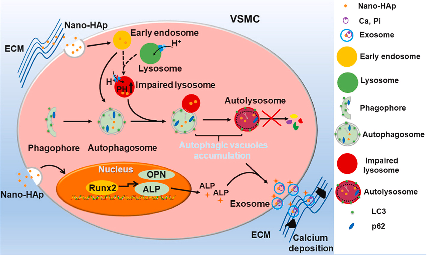
Vascular calcification (VC) is a common feature of aging, diabetes mellitus, chronic renal failure, and atherosclerosis. The basic component of the VC is hydroxyapatite (HAp). Nanometer-sized HAp (nHAp) has been identified to play an important role in the development of pathological calcification of the vasculature. However, whether nHAp can induce calcification in vivo and the mechanism of nHAp in VC progression remains unclear. We found that nHAp was present in vascular smooth muscle cells (VSMC) and their extracellular matrix (ECM) in the patient's calcified arteries. The synthetic nHAp has similar morphological and chemical properties to the native nHAp recovered from calcified arteries. The nHAp stimulates osteogenic differentiation and accelerates the mineralization of VSMC in vitro. Synthetic nHAp can also directly induce the VC in vivo. Mechanistically, ATPase is used for acidification, thus preventing the autophagic flow in VSMC. Lysosomal reacidification by cyclic 3 ′, 5 ′ -adenosine monophosphate (cAMP) significantly enhanced autophagic degradation and attenuated nHAp-induced calcification. Accumulated autophagosomes and autolysosomes are converted into calcium-containing exosomes, secreted into the ECM and accelerate vascular calcium deposition. Inhibition of exosome release in VSMC reduced calcium deposition. In summary, our results demonstrate an inhibitory effect of nHAp on lysosomal acidification, which suppresses autophagic degradation and promotes the conversion of accumulated autophagic vacuoles into exosomes, Pi and ALP loaded with unresolved nHApCa 2 +. These exosomes bud from the plasma membrane, deposit in the ECM, and form calcium nodules. Thus, nHAP accelerates vascular calcification by blocking autophagic flow in VSMC.
18915694570
Previous: BK Engineering Bacteri
Next: Revascularization-β-HS
