

Daily Bone Tumor Idea: Black Phosphorus Surface-Engineered Scaffold for Tumor Ablation and Bone Repair
(1) Abstract:
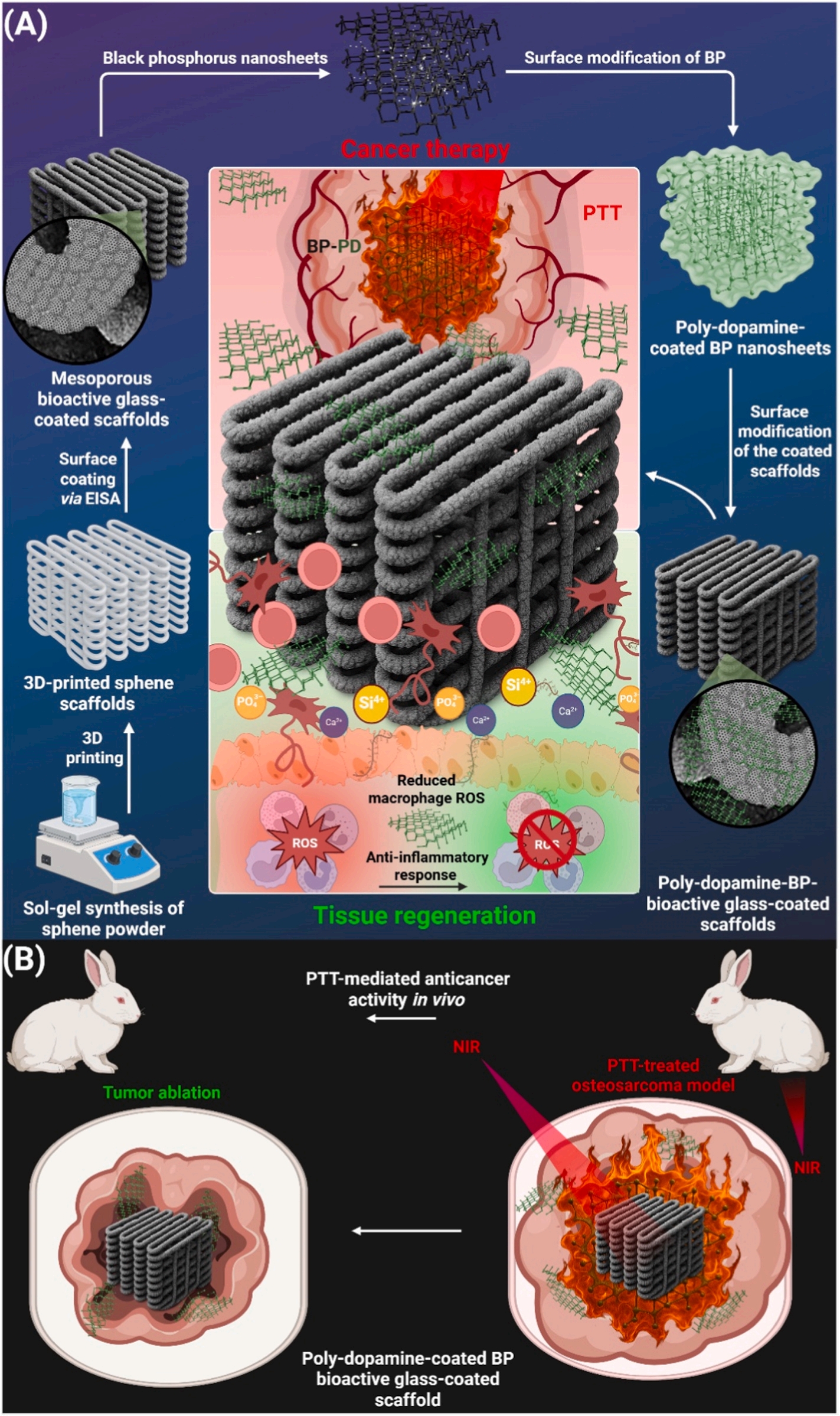
This study constructs a hierarchically surface-engineered 3D-printed sphene (CaTiSiO3) porous scaffold sequentially modified with an ordered mesoporous bioactive glass (OMBG) coating and a black phosphorus–polydopamine (BP-PD) nanocomposite layer, aiming to combine residual osteosarcoma cell elimination with repair of tumor-associated bone defects. OMBG promotes calcium phosphate deposition and osteogenesis-related responses and provides near-infrared heating; BP-PD further enhances photothermal response and tumor cell ablation. In vitro, the scaffold supports human mesenchymal stem cell osteogenesis and suppresses osteosarcoma cells. In a rabbit distal femoral VX2 model, it suppresses local tumor growth, invasion, and distant metastasis; imaging shows reduced tumor, edema, and intramedullary invasion, with preservation of peri-implant bone and no obvious systemic toxicity.
(2) Background:
Bone tumor resection often creates large bone defects, and residual malignant cells coexist with the need for bone regeneration, making single-treatment approaches insufficient. 3D-printed porous ceramic scaffolds provide structural support but require surface functionalization to confer antitumor and pro-osteogenic activity. Black phosphorus has near-infrared absorption and osteogenic degradation products but limited stability; polydopamine can improve stability, adhesion, and photothermal performance.
(3) Main Content:
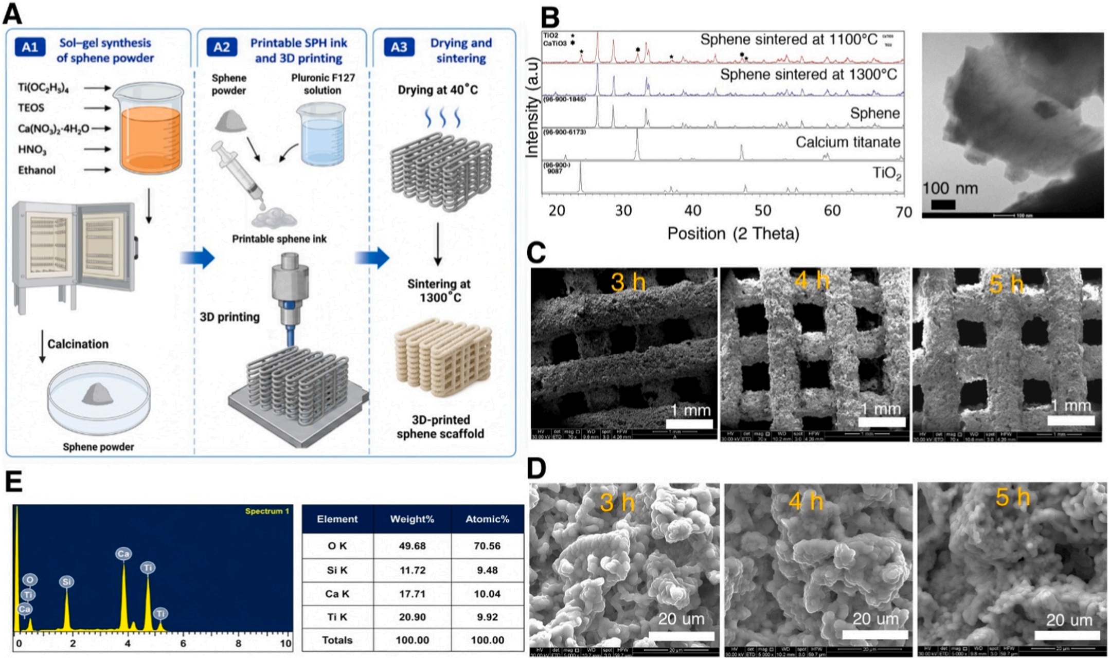
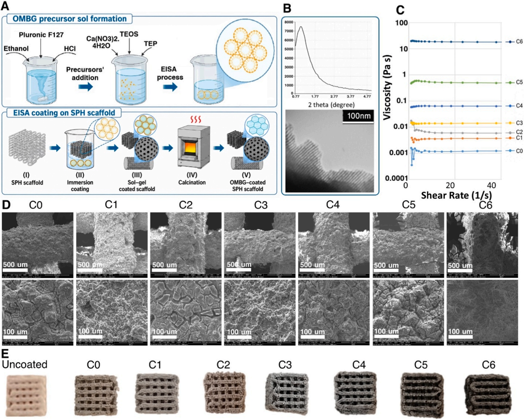
Sphene powder was prepared by sol–gel, followed by 3D printing and sintering to obtain porous scaffolds. An OMBG coating was constructed by evaporation-induced self-assembly, and BP-PD was deposited by in situ dopamine polymerization. OMBG improves surface bioactivity, ion exchange, and mineralization; BP-PD increases light absorption, surface roughness, and photothermal conversion. In vitro evaluations included simulated body fluid mineralization, ion release, photothermal heating, osteosarcoma cell viability, mesenchymal stem cell adhesion and osteogenic markers, and macrophage anti-inflammatory and antioxidant responses. In vivo, a rabbit distal femoral VX2 model received near-infrared irradiation, with DR, MRI, CT fusion reconstruction, and histology used to assess tumor control, bone structure, metastasis, and biosafety.
(4) Study Design:
The study combined material construction with in vitro and in vivo evaluation. Material characterization covered phase, morphology, elemental distribution, Raman, surface topography and roughness, compressive properties, mineralization, and ion release. Cell experiments used osteosarcoma cells, human mesenchymal stem cells, and macrophages. Animal experiments included sham, control, and treatment groups, with sampling at multiple time points to evaluate tumor burden, invasion depth, distant metastasis, and implant stability. The model was an early-intervention model, VX2 is a transplantable carcinoma rather than osteosarcoma, and the observation period was relatively short, as noted in the original article.
(5) Results:
Sequential OMBG and BP-PD modification did not obviously compromise overall scaffold compressive performance. OMBG promoted early calcium phosphate deposition, and BP-PD produced a surface more favorable for mineral layer maturation. Under near-infrared irradiation, BP-PD-modified scaffolds showed greater heating and stronger photothermal killing of osteosarcoma cells; cytotoxicity without irradiation was low. Scaffold-conditioned medium supported mesenchymal stem cell viability, type I collagen, and osteocalcin expression, and promoted anti-inflammatory and antioxidant macrophage phenotypes. In vivo, the treatment group showed lower tumor volume, peri-tumoral edema, and intramedullary invasion, reduced local infiltration and distant metastasis, better preservation of peri-implant bone structure, and no obvious pathological abnormalities in major organs.
(6) Future Directions:
This work integrates surface engineering, photothermal antitumor action, and pro-osteogenic cues into a single implant, but osteogenic evidence is mainly in vitro, and in vivo new bone regeneration requires quantification. Future work should extend observation, evaluate long-term degradation and mechanical retention, optimize BP stability, coating uniformity, and immune response, dissect tumor–bone interface mechanisms, validate in models closer to osteosarcoma biology and different defect severities, and distinguish the independent contributions of each functional layer.
We provide full-process services for various material designs (including highly innovative scheme design, preparation, characterization, in vitro and in vivo validation (cell/animal experiments), etc.): a postdoctoral team (with multiple high-impact publications in Zone 1 journals) can meet all kinds of tricky and challenging requirements, including hydrogels / drug delivery / nanomaterials / polymers / liposomes / microneedle drug delivery, etc. We can assist with project design / NSFC grant applications / publication of high-impact Zone 1 and Zone 2 papers in journals such as AFM, SCI, AM, etc. Welcome to consult 👇️
17768014569 (Senior Engineer Gao)
17761925168 (Engineer Zhang)
18013729810 (Engineer Yang)
18915694570
Previous: Nanomaterial Customiza
Next: Custom Nanomaterial Se


